Doppler Mode
Doppler assess a frequency change in the returning ultrasound signal. If a signal impacts a moving object; it returns to the transducer at a different frequency.
- Movement towards the transducer INCREASES the frequency of the return signal
- Movement away from the transducer DECREASES the frequency of the return signal
- The amount of Doppler shift is related to the velocity of movement (higher velocity flow; more Doppler shift)
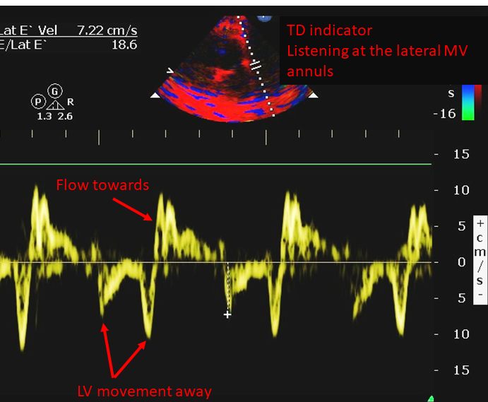
- In “regular” Doppler, the moving object is blood flowing. However, in tissue Doppler Imaging the moving object is the tissue itself
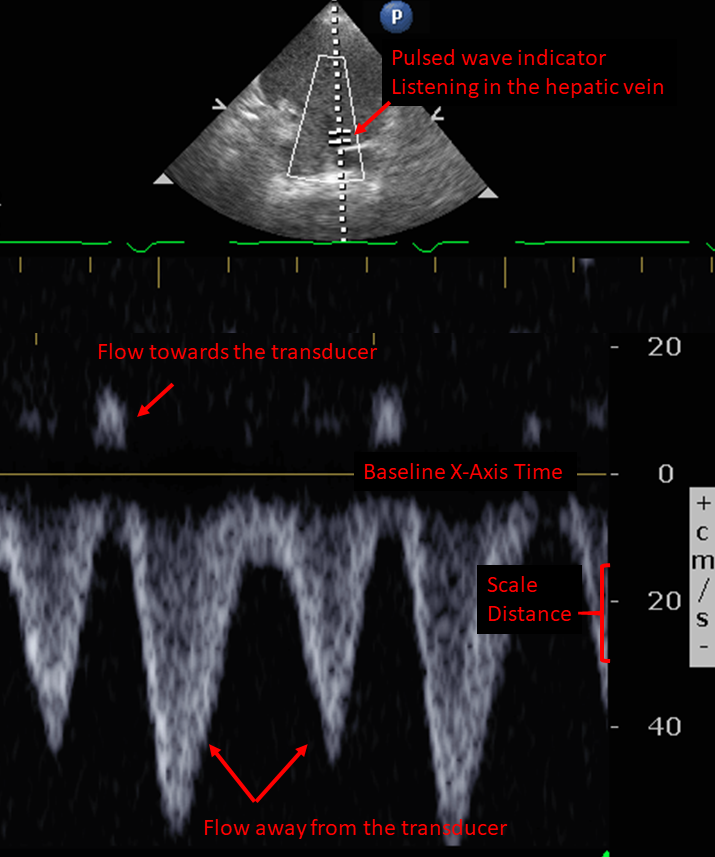
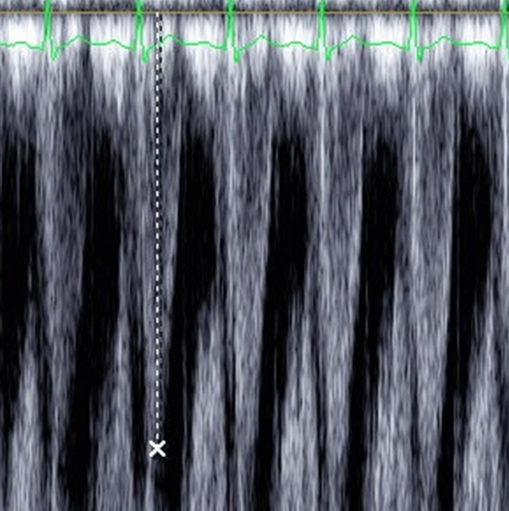
- Pulsed signal allows listening at a specific point. However, it will alias at high flows (Figure 2)
- A grey value (white to black) is assigned to the Doppler shift
- Along a Time (sec) X-axis vs. Distance (cm, or m) Y-axis to display velocity (cm/sec)
- X-axis is called the baseline
- Used to assess stroke volume and hepatic and portal venous flow (Figure 1)
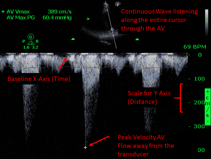
- One crystal continuously sends and another on one receives the signal
- It listens along the entire cursor, not a specific spot, and it does not alias.
- It measures high flow jets
- A grey value (white to black) is assigned to the Doppler shift
- Displayed on distance vs time axis
- Used to assess for elevated systolic pulmonary artery pressure, and to grade/assess aortic stenosis (Figure 3)
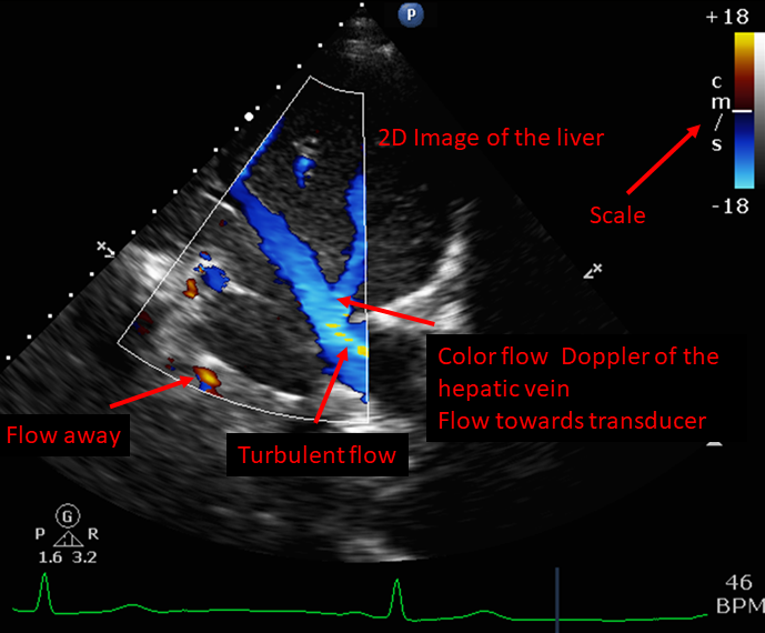
- Type of pulsed wave Doppler (will alias at high flows)
- A color value is assigned to positive and negative Doppler shift
- The CFD is overlaid onto a 2D image to create Duplex Doppler
- Used to identify location of normal blood flow, identify pathologic blood flow (i.e. ventricular septal defect), and assess valvular function
- Type of pulsed wave Doppler
- Measures tissue movement
- Most commonly the left ventricle at the mitral annulus
- Used to asses diastolic function
In color Doppler, by convention flow towards the transducer is red, flow away is blue. This is not related to arterial or venous flow
Figure 1 - Pulsed Wave Doppler
Figure 2 - Aliasing at high flows