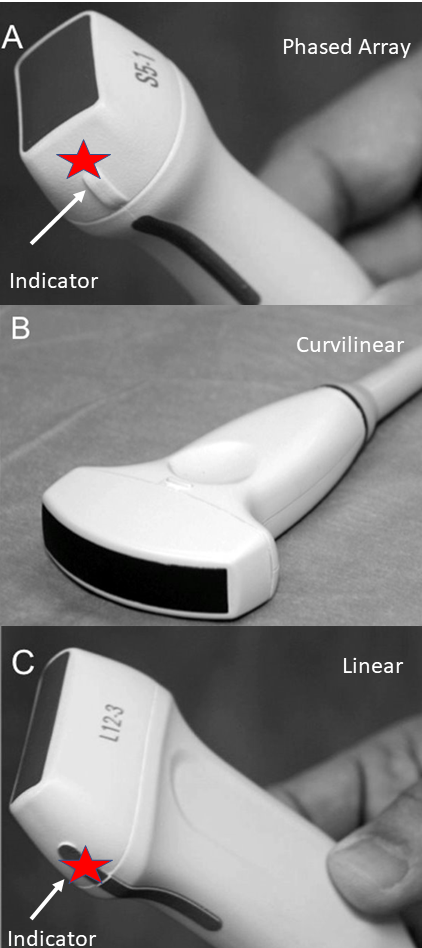
Probe Selection
Lung imaging focuses on the pleura and pleural interface. The linear probe (Figure 1) is best used to assess the pleura and the pleural interface. The Curvilinear (or phased array) is used to assess artifacts, B-lines, and A-lines. Both probes can show lung sliding (or no sliding), and this is used to diagnose pneumothorax
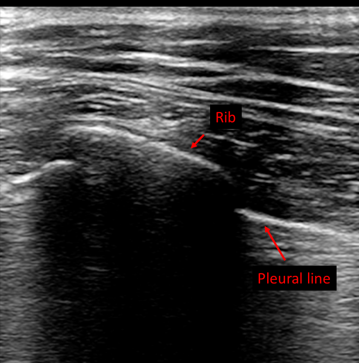
- High frequency, and hence best for superficial imaging
- Excellent visualization of the pleura
- Assess pleural abnormalities
- Assess lung sliding
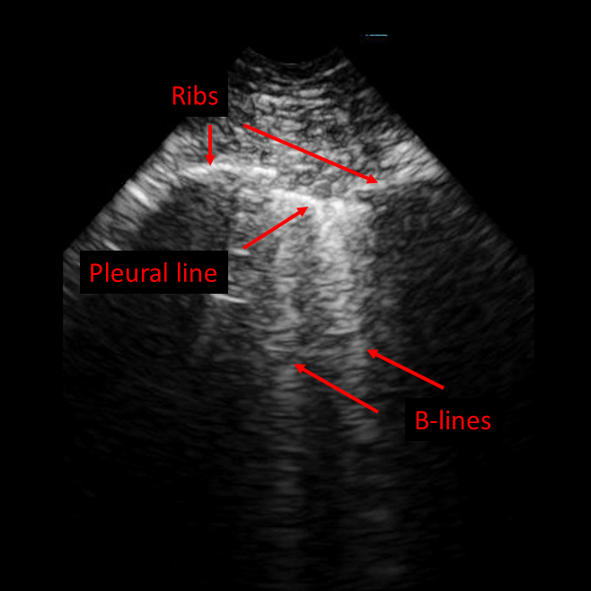
- Low frequency, and hence can be used for deep lung imaging (Figure 2)
- See several rib spaces
- Able to see B-lines and A-lines
- Able to see effusion and collapse
- Used in some centers, and acceptable if using for other imaging
- Does not allow multiple ribs spaces
- Most systems have a lung preset/exam type
- Can also use abdominal/FAST presets. However, will need to optimize gain and depth