Inferior Vena Cava
The ideal IVC view is longitudinal allowing visualization as it courses through the liver and enters into the right atrium.
- Once the right ventricle and atrium are identified in the subxyphiod
- Rotate the probe counterclockwise 90 degrees
- Longitudinal images are best obtained for IVC measurements (Clip 1)
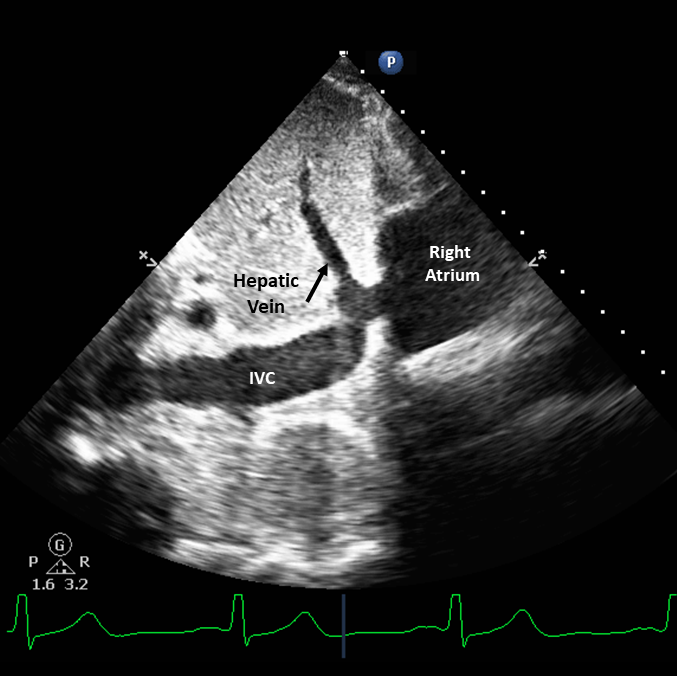
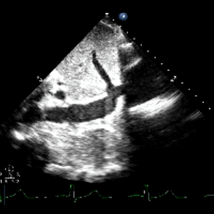
- Look for the confluence of the hepatic vein into the IVC (Figure 2)
- The IVC can be differentiated from the aorta by its thinner walls and collapse during respirations
- May need to increase the depth
- IVC longitudinal view, indicator is towards the patient’s head
- Indicator on the right of the screen
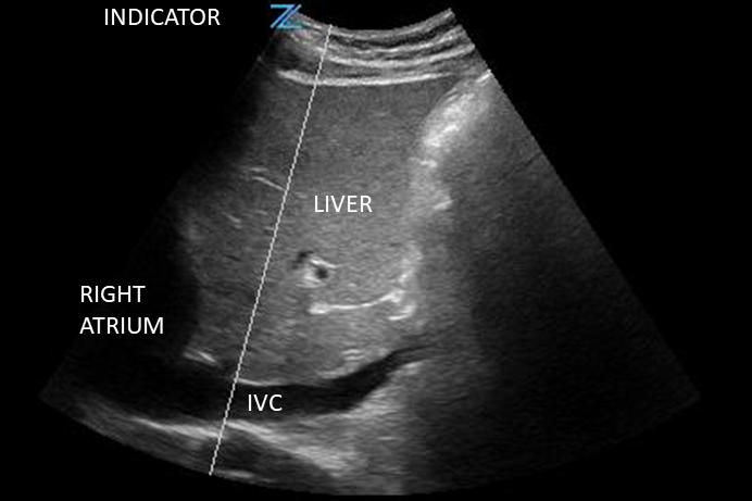
- IVC the indicator is towards the patient’s head
- Indicator on the left of the screen
- This view allows assessment for IVC diameter, and IVC diameter change with respiration
- M-mode or 2D caliper can be used to make the measurements
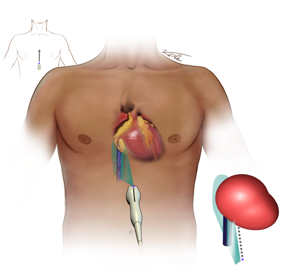
Figure 1 - Transducer Placement
Figure 3 - Ideal IVC - Cardiac Preset
Figure 4 - Ideal IVC - Abdominal Preset